TRI microscopy facility: Equipment and services
 |
Brightfield microscopy: Nikon Brightfield (rm4026): Objectives: 4X, 10X, 20X,40X, 60X and 100X Oil objectives. phase contrast condenser.NIS Basic Elements Software.DS Fi1 colour camera. Manual operation. Application: For slides only. For imaging unstained samples (phase contrast). For H&E, DAB and other color staining. |
|
|
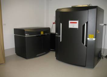
Slide Scanning: Application: For slides only. For imaging whole tissue sections and the ability to zoom to the single cell level. Slide preparation crucial! Slides must be clean and preferably coverslipped on an automatic coverslipper (contact facility manager for further information or click here for more info) |
|
|
Epifluorescence Microscopy: CellSens Software, inverted IX73 microscope. DP73 colour (pixel shift) camera (room 4035) OR XM10 monochrome camera (Room 4066, 5026 and 6067) |
|
|
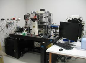
Olympus Motorized upright Fluorescence microscopes (rm 4026) 10X, 20X and 40X UPlanSAPO, 60X and 100X Oil UPlanSAPO (correction collar) objectives. Motorised DIC condenser CellSens Software, unright BX63F microscope. DP80 dual chip colour/monochrome (pixel shift) camera. Motorized X/Y, objective turret and filter turret X-Cite LED fluorescence light source, multiple filters: 350 DAPI, 470 GFP, 560 Cy3, 650 Cy5 Application: Excellent for fluorescence scanning of tissue sections or high resolution epifluorescence imaging. |
|
|
Confocal microscopy: Zen Software. 4 Lasers: Argon/2 (458,477,488,514), DPSS 561-10, HeNe633 and Diode 405-30. One spectral and 2 filter based PMT channels, one transmitted light detector. Filter cubes for visual observation GFP, DsRed and DAPI. Application: For high resolution, confocal imaging, Z-stacks, Mosaic, FRET, FRAP. Versatile, high-end imaging. For colocalisation, resolving power 250-350nm in X/Y and 500-700nm in Z (for higher resolution, please see OMX-Blaze 3D-SIM superresolution microscope below) |
 |
Olympus FV1200 Confocal Microscope 20X UPLSAPO, 60X UPLSAPO and 100X UPLSAPO Oil objectives, 10X UPLNSAPO, 20X UPLSAPO and 60X LUCPLFLN air. DIC and phase. FV10 Software. 5 solid state Lasers: LD405nm (50nw), LD473 (15mw), LD559 (15mw), LD635nm (20nw) and now LD748nm. 2 channel high sensitivity GaSP detectors, 2 spectral, 1 filter based PMT and one transmitted light detector. Filter cubes for visual observation U-FUW Ex: 340-390nm Em: 420nm long pass. U-FBVW Ex: 400-440nm Em: 460nm long pass. U-FBN Ex: 470-495nm Em: 505nm long pass U-FGW Ex: 530-550nm Em: 570nm long pass Application: For high resolution, confocal imaging, Z-stacks, Mosaic, high-end imaging with additional features including HDRi, amazing spectral unmixing ability. Background reduction, high sensitivity GaSP detectors. resolving power 250-350nm in X/Y and 500-700nm in Z (for higher resolution, please see OMX-Blaze 3D-SIM superresolution microscope below) |
|
|
Live Cell Imaging Axiovision Software. AxioCam MRm colour camera. Motorized X/Y stage, temperature and CO2 control.Mercury lamp, filters for GFP, RFP…and others Application: For long term live imaging in brightfield and fluorescence (up to 5 days!). Good for imaging plastic multiwell dishes, or multiwell slide format. Long working distance, low magnification objectives for imaging large number of cells rather than single cells (see Spinning disc confocal for high resolution live imaging of single of single cells and subcellular features below) |
 |
Olympus CellR Live Cell Imager: 10X, 20X,40X UPlanSApo and 60X Oil UPlanSApo objectives. DIC motorized condenser Xcellence Software. Hamamatsu Orca Flash 2.8 camera. Motorized X/Y stage, temperature and CO2 control. LED light engine, multiple filters: 350 DAPI, 470 GFP, 485 FITC, 556 RFP, 560 Cy3, 650 Cy5 Application: For long term live imaging in brightfield and fluorescence (up to 5 days!). best for imaging glass/optical plastic bottom multiwell slides. Long working distance, low magnification objectives as well as better high magnification oil objectives for imaging smaller number of cells (see Spinning disc confocal for high resolution live imaging of single cells and subcellular features below) |
|
High end imaging |
|
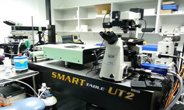 |
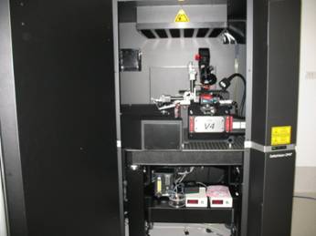
Multiphoton (rm1006) Animal house: Peripheral equipment include oral anaesthetic for mice, warming blanket and monitoring equipment. Heated chamber as well as heated deep chamber to accommodate whole ex-vivo organs with gas perfusion including CO2 and/oxygen.] Access is limited by facility manager and prior arrangements made for use. Application: Deep imaging into samples (up to 2mm), Live imaging of mice or ex-vivo organs at the cellular level. Other samples. Wide variety of dyes and fluorophores can be imaged. See list of filters and dichroic mirrors available here. |
|
 |
OMX Blaze deconvolution structured illumination (SIM) superresolution microscope (rm 4026): What is it?: OMX-Blaze 3D-SIM (Structured illumination microscopy) super-resolution microscope. Uses a frequency based interference pattern, unique deconvolution algorithms, surprisingly simple to use platform with 3 available laser lines, 405nm, 568nm and 488nmw and 2 sCMOS cameras. For two-fold improvement on Confocal resolution in x,y and z. VERY fast! Features: 2 X sCMOS Cameras Custom liquid cooled, 15 bit, 5.5 Megapixel chip, 6.45 um pixels (3D-SIM operation at 512 x 512, Widefield operation 1024 x 1024) Must be matched to 60x objective for SI imaging Dedicated computer with fast drive architecture, camera interface board and cabling. 3 lasers: 405, 488, 546nm Solid state. Application: When better resolution is needed to discern colocalisation or to resolve fine structures than that seen in confocal microscopy 90-130nm in X/Y compared to 180-250nm X/Y confocal 250-350nm in Z compared to 500-700nm Z in confocal For sample preparation details click here. |
|
Single Focal adhesion dynamics imaged on our system by Samantha Stehbens |
Nikon/Spectral Spinning Disc Confocal microscope: Cameras: FAST: Photometics Evolve Delta 512 X 512, EMCCD, 16 X 16um pixels, fast 33.7fps full resolution, 224 at 64X64, 16 bit High Resolution: Andor Clara CCD Camera 1392 x 1040 , -55°C cooling, 6.45 x 6.45 pixel size , 11fps frame rate, 16 bit Application: For live imaging of intracellular processes such as vesicular, transport, receptor endocytosis, microtubule dynamics, cell adhesion and movement in A QUANTIFYABLE manner! NOTE: SDC usage is to be conducted by collaboration with Dr Samantha Stehbens . SDC is technically challenging and prior consultation is essential. |
 |
Analysis Workstations What do we have?: Software packages we use (in order of complexity): Image J- Free, easy, useful for simple analysis, robust NIS-Elements- Nikon software, user friendly, versatile Visisopharm- Analysis of whole tissue sections on a slide, can count cells, staining, whatever you teach it to count…requires some time to learn to use Imaris- For 4D- X/Y/Z and time images acquired on any of theinstruments described. Can quantitate, make movies, etc… just about anything. requires some time to learn to use, online tutorials available. PLEASE NOTE: We aim to help, but customized analysis takes time to work out, we are happy to assist, but you are required to put in the time for your own analysis as well! |


