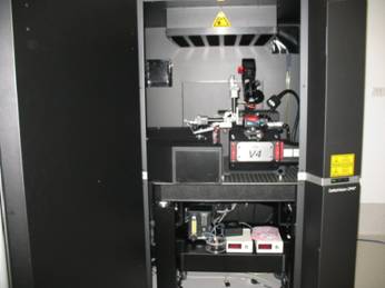
TRI Microscopy core facility
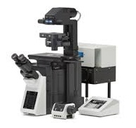
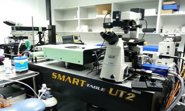
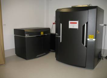
The TRI has an outstanding microscopy facility including inverted fluorescence microscopes on most research floors and a fully equiped facility room of high end instruments. For analysis purposes, computers containing a range of analysis software are also available for use.
The facility staff provides researchers with the training and knowledge to efficiently generate high quality reproducible images.
The facility is open to all users including external and commercial with the same charge rate.
CHARGES AND TERMS OF USE
To enquire about facility charges, please email [email protected] or access the PPMS booking system.
![]()
EQUIPMENT AND SERVICES
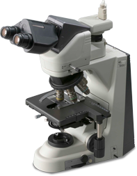 |
Nikon Eclipse 50i Brightfield Microscope Upright Manual Facility room 4026 |
Objectives: 4X, 10X, 20X, 40X, 60X and 100X Oil objectives Camera/Detector: DS-Fi1 colour camera Light Source: Halogen lamp for brightfield Software: NIS-Elements Basic Research Application: For slides only e.g. H&E, DAB and other histological stains |
|
|
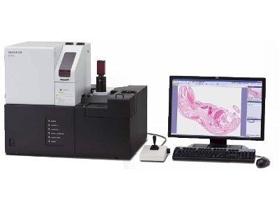
Olympus VS120 Slidescanner Microscope Upright Fully automated Facility room 4026 |
Objectives: 2X (overview), 10X, 20X or 40X for actual scanning. Polarization also available Camera/Detector: Allied Pike 505 colour camera Light Source: Halogen lamp for brightfield Software: VS-ASW Application: For batch scanning up to 100 histologically stained slides. Service run by facility |
|
|
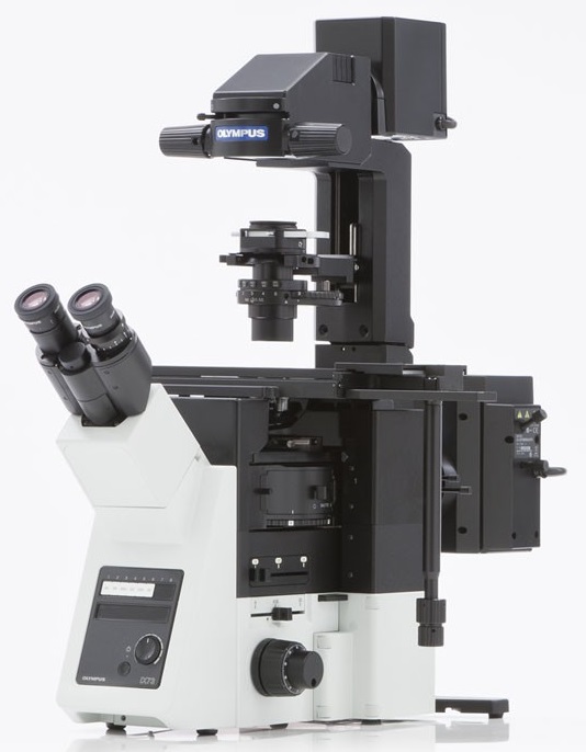
Olympus IX73 Microscope Inverted Manual Rooms 4035, 4066, 5026 and 6067 |
Objectives: 2X, 4X, 10X, 20X, 40X, 60X Oil Camera/Detector: XM10 monochrome camera (room 4066, 5026 and 6067). DP73 colour camera in room 4035 Light Source: Halogen lamp for brightfield. Metal halide lamp for fluoresence (DAPI, GFP, CY3, CY5). Software: Cell Sense Application: For checking slides, dishes, plates, staining quality, transfection efficiency and taking simple images |
|
|
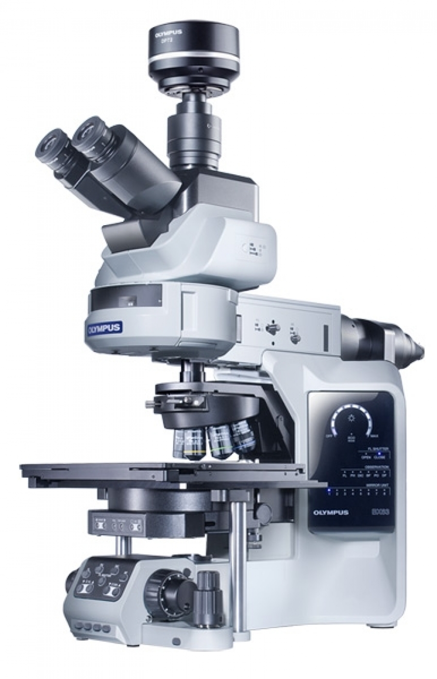
Olympus BX63 Microscope Upright Motorised Facility room 4026 |
Objectives: 4X, 10X, 20X and 40X, 60X Oil and 100X Oil Camera/Detector: DP80 monochrome/colour camera Light Source: Halogen lamp for brightfield. X-Cite LED for fluoresence (DAPI, GFP, CY3, CY5) Software: Cell Sense Application: Slides only, automated imaging of multiple colours and single slide tile scanning |
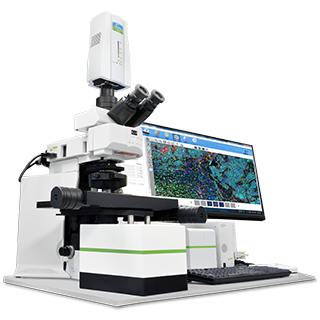 |
PerkinElmer Vectra III Spectral Scanner Microscope Upright Automated Facility room 4026 |
Objectives: 4X, 10X, 20X and 40X Camera/Detector: PerkinElmer Spectral camera Light Source: Halogen lamp for brightfield. Metal halide lamp for fluoresence (DAPI, GFP, CY3, CY5). Software: Vectra, Phenochart, Inform Application: Slides only, automated imaging of up to 6 slides, H&E, IHC, IF, OPAL stains. Spectral detection and separation of overlapping biomarkers. |
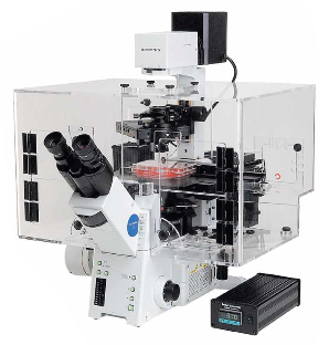 |
Olympus IX81 Live imaging Microscope Inverted Motorised Room 4024 |
Objectives: 1.25X, 2X, 4X, 10X, 20X, 40X, 60X Oil Camera/Detector: Hamamatsu Orca Flash 2.8 monochrome camera Light Source: Halogen lamp for brightfield. Metal halide lamp for fluoresence (DAPI, GFP, CY3, CY5) Software: Cell Sense Application: Slides, dishes, plates. For long term fast live imaging of cells/spheroids, etc. in multiple fluorescent colours plus brightfield, phase or DIC. Temperature and gas controlled. |
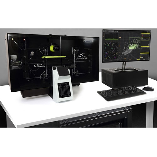 |
Phasefocus Livecyte Live imaging Microscope Inverted Automated Room 4024 |
Objectives: 4X (preview), 10X, 20X and 40X imaging Camera/Detector: Monochrome sCMOS camera Light Source: LED for brightfield and fluoresence (DAPI, GFP, CY3), 650nm laser for quantitative phase imaging Software: Cell Analysis Toolbox and associated APPs Application: Slides, dishes, plates. For long term ptychography and fluorescence live imaging of cells. Fully automated focusing and imaging. Adjustable QPI field of view. Temperature and gas controlled. Dedicated data processing unit and network storage. |
|
|
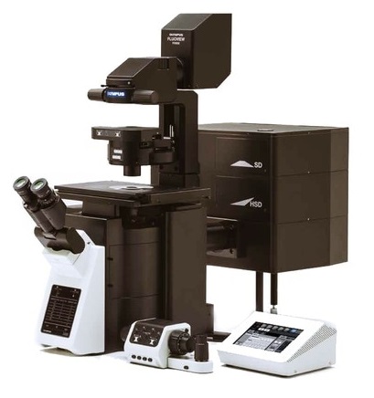
Olympus FV1200 Confocal Microscope Inverted Motorised Facility room 4026 |
Objectives: 4X, 10X, 20X, 40X, 40X Oil, 60X Oil and 100X Oil Camera/Detector: 2 spectral and 1 filter PMT, 2 GaASP PMT and 1 transmitted light detector Light Source: Halogen lamp for brightfield, metal halide lamp for fluoresence (DAPI, GFP, CY3). Lasers: 405, 473, 559, 635 and 748nm Software: FluoView 10 ASW Application: Slides, dishes, plates. For high resolution confocal imaging, Z-stacks, Mosaic, high-end imaging. Resolving power 250-350nm in X/Y and 500-700nm in Z |
 |
Olympus FV3000 Confocal Microscope Inverted Motorised Facility room 4026 |
Objectives: 4X, 10X, 20X, 40X, 40X Oil, 60X Oil and 100X Oil, 30X and 60X Silicone Oil Camera/Detector: 4 GaASP spectral PMTs and 1 transmitted light detector Light Source: LED for brightfield, metal halide lamp for fluoresence (DAPI, GFP, CY3). Lasers: 405, 445, 488, 514, 561, 594 and 640nm Software: FluoView 31S Application: Slides, dishes, plates. For high resolution, confocal imaging, Z-stacks, Mosaic, high-end imaging. Resolving power 250-350nm in X/Y and 500-700nm in Z. Includes a hybrid resonant/galvonometer scanner for faster imaging and a stagetop incubator |
 |
Nikon/Spectral Spinning Disc Confocal Microscope Inverted Motorised Facility room 4026 |
Objectives: 4X, 10X, 20X, 40X, 40X Oil, 60X Oil and 100X Oil Camera/Detector: Andor Clara CCD camera 1392 x 1040, 6.45x6.45um pixel size , 11fps. Photometics Evolve Delta EMCCD camera 512 X 512, 16X16um pixel size, 33.7fps. Light Source: Halogen lamp for brightfield, LED for fluoresence (DAPI, GFP, CY3). Lasers: 405, 445, 514, 488, 561 and 640nm. Software: NIS-Elements Advanced Research Application: Slides, dishes, plates. For high resolution, fast, confocal imaging, Z-stacks, Mosaic, high resolution intra cellular live imaging. Temperature and gas controlled. 355nm laser ablation system from Roper, TIRF imaging |
|
|
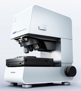
Olympus LEXT OLS 4100 Microscope Upright Motorised Facility room 4026 |
Objectives: 5X, 10X, 20X, 50X, 50X Long Working Distance, 100X Camera/Detector: High sensitivity PMT Light Source: 405nm laser Software: OLS LEXT Software Application: For measuring surface features of materials up to 10nm resolution |
|
|
Multiphoton Microscope Upright and Inverted Motorised Facility room 4026 |
Objectives: 16X, 40X and 20X Camera/Detector: 4 High sensitivity PMTs Light Source: Tunable emission laser 690-1040nm. OPO included to modulate output wavelength to IR spectrum (Optical Parametric Oscillator) Software: LaVision Biotek software, Imspector and Imaris on floating license Application: Deep imaging into samples (up to 2mm), Live imaging of mice or ex-vivo organs at the cellular level. Wide variety of dyes and fluorophores can be imaged. Supports two Nikon microscopes: Eclipse Ti-U inverted and Eclipse FN1 upright microscopes. Run by a Spectra Physics Ti:Sapphire pulsing. The Inverted microscope has a fully motorized X/Y/Z stage. Has a 64 beam splitter attached as well as high speed camera. Options also include 5 PMTs (including one ultrasensitive) and a selection of filters for flexibility. The upright microscope is fitted with a Z-motorised intravital stage, 4 PMTs (including ultrasensitive), and a wide selection of filters. Peripheral equipment include oral anaesthetic for mice, warming blanket and monitoring equipment. Heated chamber as well as heated deep chamber to accommodate whole ex-vivo organs with gas perfusion including CO2 and/oxygen. Access is limited by facility manager and prior arrangements made for use. |
|
|
GE Healthcare OMX Blaze 3D Structured Illumination Microscope (SIM) Inverted Motorised Facility room 4026 |
Objectives: 60X Oil Camera/Detector: 2 sCMOS Cameras Custom liquid cooled, 15 bit, 5.5 Megapixel chip, 6.45 um pixels (3D-SIM operation at 512 x 512, Widefield operation 1024 x 1024) Light Source: 405, 488 and 568nm lasers Software: Deltavision OMX for acquisition and a separate reconstruction computer with SoftWoRx Application: Structured illumination super-resolution microscope. Uses a frequency based interference pattern, unique deconvolution algorithms. When better resolution is needed to discern colocalisation or to resolve fine structures than that seen in confocal microscopy 90-130nm in X/Y compared to 180-250nm X/Y confocal 250-350nm in Z compared to 500-700nm Z in confocal |
|
Analysis Software 4 physical computers + 2 virtual machines Free software: |
||
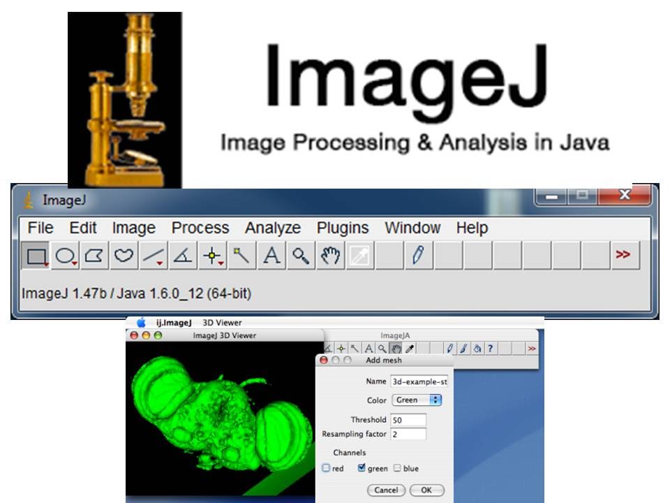 |
Image J / Fiji |
Freely available, open sourced, java based image processing software. Great for image processing and analysis with many useful plugins e.g. Bio-Formats Importer for opening proprietary files. Please contact a facility staff member for assistance with your ImageJ analysis and automated scripting. |
 |
OlyVIA |
Freely available viewing software for Olympus files (VSI, OIF, OIR, etc.) including the virtual slide files from the VS120 slide scanner, timelapse data from the IX81 live imaging microscope, etc. Please ensure you are using the latest version which includes a "Crop and Export" function. |
 |
FV10 Viewer / FV31S Viewer | Freely available viewing software for the FV1200 OIF files and the FV3000 OIR files |
 |
NIS-Elements Viewer | Freely available software for viewing Nikon ND2 files |
|
Licensed Software: |
||
 |
NIS-Elements Advanced Research | Nikon's acquisition and analysis software. Powerful software capable of image segmentation, measurements, tracking, morphology, counting, etc. |
| Visiopharm | Automated trainable analysis software for batch whole section/slide analysis. | |
 |
Cell Sense | Olympus's acquisition and analysis software including Olympus's deconvolution. |
 |
Phenochart/Inform | PerkinElmer's image analysis and channel unmixing software for the spectral images from the Vectra 3.0 microscope. |
 |
Spotfire | Tibco's data analysis software used mainly for extracting data genereated from Inform. |
 |
Cell Analysis Toolbox + associated APPs | Phasefocus's analysis software for quantifying the QPI timelapse data acquired on the Livecyte microscope. Virtual machine access via remote connection. |
 |
Imaris | Bitplane's powerful analysis software. Can open almost every proprietary file type and great at opening large files. Can view and render 3D datasets, automated tracking of cells from live imaging datasets. |
![]()
Contact the Microscopy Facility
For more information or bookings please:
If you cannot book the instrument you want:
Instruments cannot be booked until a certificate has been issued, which will only occur after training has been completed. The certificate represents your level of training/expertise on a particular instrument. Contact the TRI Microscopy team for further information on ext: 37772 or email m[email protected]
![]()
Microscopy Club
The Microscopy Club launched in September 2017 to bring together the collective knowledge and experience of those using the Microscopy Facility at TRI. See what was covered at the first Microscopy Club meeting here or stay tuned to the weekly events email for details about upcoming sessions.


