Medical research to benefit from TRI imaging boost
TRI will join a gathering of global imaging experts in Denmark, exploring ways 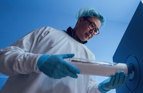 to combine technologies that will boost medical research in areas including
cancer, immunology, bone and tissue regeneration.
Senior Preclinical Imaging Scientist Dr Brian Tse from TRI core facility
Preclinical Imaging will be one of 24 experts to take part in the Molecule to Human Boot Camp in September.
He says TRI Preclinical Imaging has about 100 different users each year, including up to 40 first-time users, working across cancer biology, immunity, therapeutics, heart disease, bone development and tissue regeneration.
“I want to use the boot camp to learn how imaging at the large scale – that is, preclinical imaging – and small scale – light microscopy – can be combined to best effect,” Dr Tse says.
“There is so much overlap in the technology shared between preclinical imaging and microscopy in data acquisition, analysis and visualisation.
“It’s an incredible amount to learn and my aim is to speak the language of both microscopy and preclinical imaging, foster collaborations across both areas, and share the knowledge at TRI to enhance research quality and outcomes.”
Preclinical Imaging has technology including:
- microCT: commonly used to study bone diseases such as osteoporosis or new bone formation
- microPET-CT: for tumour detection, assessment of response to therapy and cancer metabolism studies
- high frequency ultrasound: to assess tissue vascularisation and heart function
- photo acoustics: for detection of diseases such as thrombosis, atherosclerosis and cancer using nanoparticles
- optical imaging: to track the location of cancer, immune cells and microbes such as bacteria and viruses
- nuclear magnetic resonance: for body composition analysis, for research into obesity and diabetes
- planar X-ray: useful to image surgical implantations and bone fractures
- gamma counting: for biodistribution analysis of injected radioactive imaging agents
- image-guided radiotherapy: commonly used for targeted irradiation of cancers of the brain, central nervous system, prostate and breast.
Recently, TRI Preclinical Imaging increased its expertise and infrastructure support for theranostics, an emerging field that combines diagnostic imaging and targeted therapy for treating diseases such as cancer.
The facility has also invested in image-guided radiotherapy capability for cancer research, replicating in the laboratory the systems and treatments used in hospitals and clinical settings.
“We promote the use of multiple imaging techniques in a single project as each provide different kinds of useful information,” Dr Tse says.
“This is feasible due to the team’s expertise in operating a range of preclinical technologies.”
Beyond medical research, TRI Preclinical Imaging also supports research across anthropology, materials science, marine biology, agriculture and climate change.
The boot camp in September is hosted by the Danish Euro-BioImaging node, fully funded by the Chan Zuckerberg Initiative and jointly organised by Global BioImaging and imaging organisations from three continents.


